


A Firm Science
The neuroscience of cognition lives on the uncomfortable boundary between soft and hard science.


👏 The Frontier Psychiatrists is a daily health-themed newsletter. Owen Muir, M.D., is the proprietor of this banana stand, but frankly, he is always much more excited to have co-authors. Today, I’m welcoming
, who writes a newsletter here on Substack also. She is a Neuroscientist and a very funny and joyful human. We met—and this is true—at a LARPing event at SXSW, about which I did a whole live/hysterical realist coverage of last year.
Sharena Rice, Ph.D.: In cognitive neuroscience, we are in the domain I call "a firm science."
Owen: This is as opposed to hard sciences, like biology, chemistry, and physics. But not quite “soft science” like economics or (gulp) psychology?
Sharena: Yep! The neuroscience of cognition is a fusion of hard and soft sciences. The search for the truth in this space often needs the objectification of subjective experiences and the subjectivization of objective data. Cognitive neuroscience is in a grey area between quantifiable certainties and the nuanced territories of perception.
Owen: Can you define “objectification” and “subjectification” for me?
Sharena: Objectification considers beings and phenomena like objects: they have no real sense of “internal reflection.” Subjectification is considering beings and phenomena as if they have their own minds. As Oliver Sacks said, “We are a combination of biology and biography" 1.
Dishing neurophysiology
Neural phenomena happen through the activity of neurons. Yet the scale of phenomena in a system matters.
Owen: What is a “neural phenomena”? What do you mean by “scale”?
Sharena: A neuron on its own could perform some computations, but this capability is vastly increased when the neuron is connected to other neurons.
At the level of tissues, some additional properties that were not seen at the level of single neurons would arise because the neurons are communicating with each other.
Neurophysiology in a dish leaves relatively little to scrutinize. At the levels of cells and tissues, the circuits are not interacting with the many inputs they would have during behavior, so studying neuronal activity is straightforward. One may apply a stimulus to one cell while recording from other cells to learn about synaptic properties—if X, then Y.

Neurophysiology can also be explored via circuit mapping. This allows us to see which brain regions connect. Working with slices of brains allows us to see a level of resolution that cannot be achieved with even the most powerful fMRIs. Techniques in brain slices can enable us to examine, at the levels of synapses, which types of neurons from which regions are “talking” with which types of neurons from other regions.
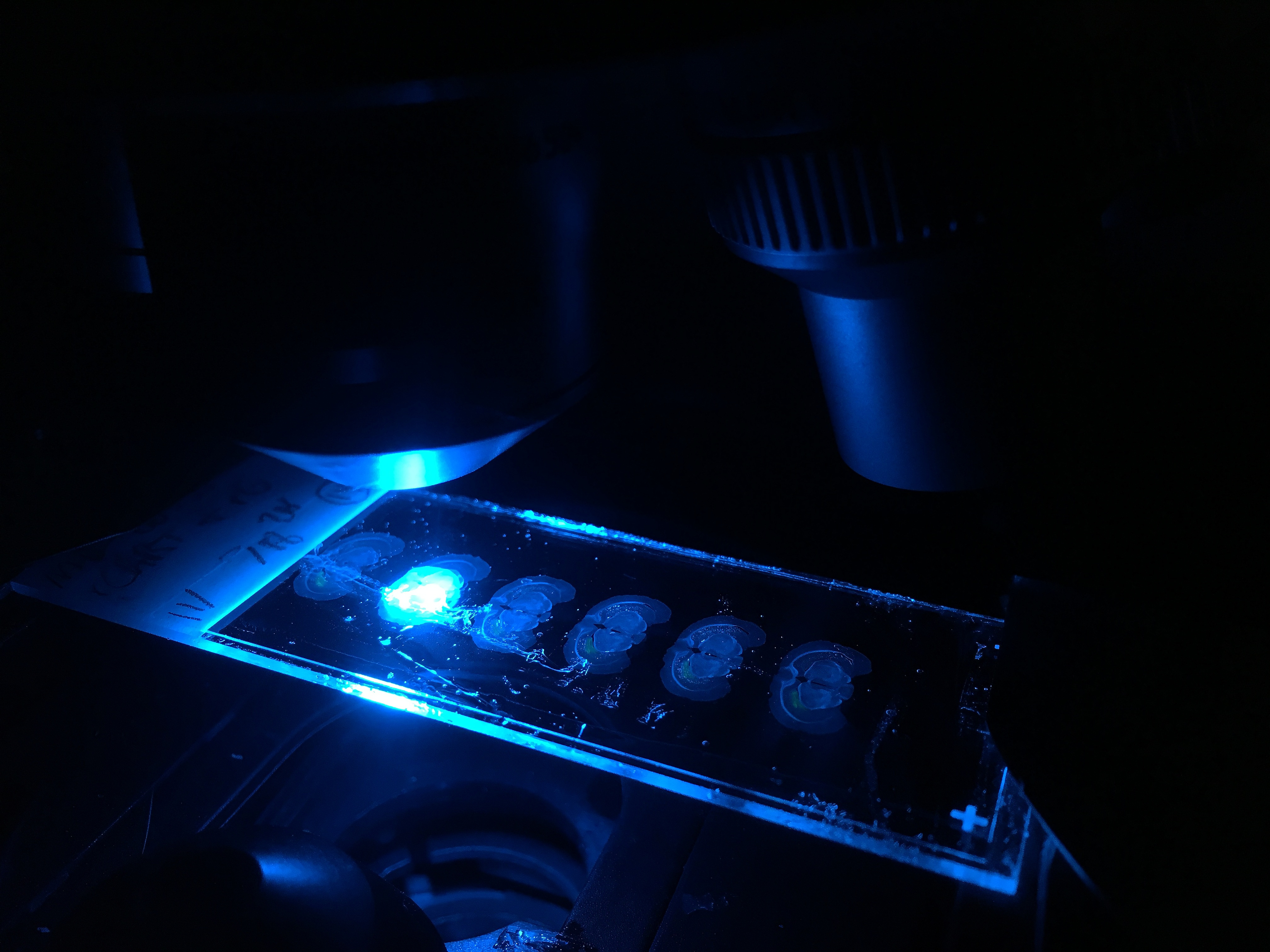
Research that involves slicing up brains or using microscopy on human brain tissue is possible after people pass away if they donate their brains for research to help future people. This information can tell about the structure, though less about the function, of the brain.
Owen: How are structure and function related?
Sharena: Structure is the physical arrangement of the brain, including its regions, circuits, and cells. Structure underpins function. Function describes what the brain is doing and how it may change with some reconstruction, such as through neuroplasticity. As an analogy, consider highways. Like the brain's structure, the structure of highways’ spans and shapes will determine what vehicles can go from one place to another. The structure of highways will not tell you about the speed, types of cars, or the amount of traffic on a given day, but the functions on these highways will.
Determining the structure and the function of the nervous system allows us to gain a sense of why and how it works.
In Vivo, IRL
Sharena: For research in living humans, research participants need to be alive to treat them adequately— and to see whether the conditions of their well-being are improving.
Owen: I’ve heard the term “In-vivo”…can you walk us through this?
Sharena: In-vivo means the subject or patient is alive. When we zoom out, from the level of organs to the level of behaving (including sleeping) mammals, there are more inputs from the outer world and more internally generated signals for the nervous system to process. Experiments are no longer as straightforward cycles of cause and effect. Instead, confounding memories and processes outside the experiments may also come into play.
Showing people the same stimulus twice does not mean they will receive or think through the information in the same ways both times. They may be preoccupied with something else one of the times and fully-attentive at another. The person may be in a different mood from scrolling through TikTok than while scrolling through LinkedIn, despite similar tasks. Scrolling through Bluesky might be more activating still, depending on how people have curated their feeds.
Good research study designs consider this: they strive to reduce confounds as much as possible within reason, have well-considered control conditions, and do their best to parse the captured signals from noise.
Opportunities and limitations with biomarkers
Owen: How will more biomarkers change this sort of research…is more coming?
Sharena: More biomarkers may emerge with advances in machine learning, computing, materials science, biosensing, and clinical trials.
In late 2023, the Nash Family Center for Advanced Circuit Therapeutics at Mount Sinai published brain biomarkers that accurately identify depressive and recovered states, track recovery trajectories, and predict relapses2. While it is impressive for a biomarker to give so much information, it involves the use of an implant for data collection from the subcallosal cingulate, the same region they implanted for deep brain stimulation to treat depression. While pain may also seem like an inherently subjective phenomenon, objective biomarker predictions for chronic pain states have been found through implants in the anterior cingulate cortex and orbitofrontal cortex3.
Owen: What is possible without implants? What can we know with non-invasive measurement tools?
Sharena: Functional MRI (fMRI) involves having a brain scan by a machine to see which brain regions become active in different tasks. Clinical research is currently ongoing to find personalized biomarkers for anxiety and depression and use these calculations to tailor a treatment using accelerated transcranial magnetic stimulation4.
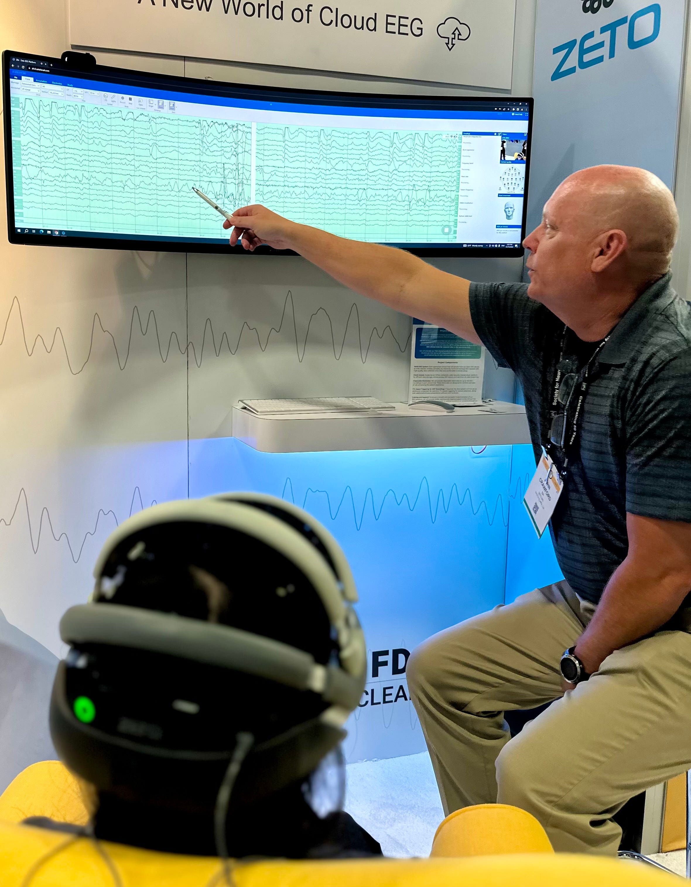
Electroencephalography (EEG) biomarkers can be helpful for noninvasively monitoring anxiety biomarkers, such as frontal alpha asymmetry5 and error-related negativity6, which are substantiated by many research studies. Other biomarkers assessed using high-density EEG (which is more precise for determining the source of the signal in the brain) can predict treatment outcomes in post-traumatic stress disorder7 and major depressive disorder8. EEG can be combined in sensor fusion with another functional near-infrared spectroscopy, a technique known as time-domain functional near-infrared spectroscopy (TD-fNIRS). TD-fNIRS has been used to measure effects on brain physiology from alcohol intoxication9 and ketamine use in humans10.

Over time, we may discover more types of objective biomarkers using combinations of sensors and less invasive techniques to give more people access to how their minds are doing. Design advances have the potential to provide biosensor systems better integration into people’s lives outside of the clinic or research lab!
While fuzzy, subjectivity is often a necessary part of the situation.
Owen: Ok, take us home!
Sharena: This brings us to the role of subjective surveys in mental health. Despite their imperfections, these surveys provide critical insights into patients' personal experiences, a realm where objective data falls short. For mental health solutions to be effective, they must embrace this amalgamation of the measurable and the experiential.
In neuroscience, particularly for those crafting solutions in mental health, recognizing the distinction between engineering for the living nervous system and engineering inanimate objects is crucial. The field requires a sophisticated understanding that respects the quantifiable while acknowledging the profound depth and variability of human experience where measurements are inaccessible or fall short.
Cognitive neuroscience, a firm science, stands as a testament to the beauty of complexity in the human mind and brain. It challenges us to find balance and harmony in the measurable and the immeasurable, a crucial understanding for anyone aspiring to innovate in the mental health space.
Thanks for reading this guest article on The Frontier Psychiatrists. Thank you
for the collab!My coverage of SXSW: Dispatch is available for your review here (where Sharena and I were thrilled to meet!):
Everything About Hype-Associated AI Risk In One Video
Disappointing Smoked Salmon Toast Consumed.
A Conflict of Interest Disclosure Regarding My SXSW Picks
Daniel Johnston had a Psychotic Illness and Lived His Dreams
Johnson and Johnson is Disrupting by Successfully Going to Market
Dear Panels on Psychedelics and Healthtech: an Insincere Apology
Psychedelic Medicine Needs to Get More Profit-Focused
Amazing Insights Regarding AI and Psychedelics (Write this Down!)
This Newsletter Files a Complaint Against Itself
An Open Letter to AI Regarding Advocating for Better DEA Regulations
Thanks for reading!
Ric Burns (Director). (2019). Oliver Sacks: His Own Life [Film]. Steeplechase Films; Vulcan Productions; Motto Pictures; American Masters Pictures; Kino Lorber. https://www.amazon.com/Oliver-Sacks-His-Own-Life/dp/B08PPXKC6N
Alagapan, S., Choi, K.S., Heisig, S. et al. Cingulate dynamics track depression recovery with deep brain stimulation. Nature 622, 130–138 (2023). https://doi.org/10.1038/s41586-023-06541-3
Shirvalkar, P., Prosky, J., Chin, G. et al. First-in-human prediction of chronic pain state using intracranial neural biomarkers. Nat Neurosci 26, 1090–1099 (2023). https://doi.org/10.1038/s41593-023-01338-z
Acacia Clinics (2024, February 11). HOPE TMS. https://www.acaciaclinics.com/hope-tms
Coan, J. A., & Allen, J. J. (2004). Frontal EEG asymmetry as a moderator and mediator of emotion. Biological psychology, 67(1-2), 7-50.
Weinberg, A., Klein, D. N., & Hajcak, G. (2012). Increased error-related brain activity distinguishes generalized anxiety disorder with and without comorbid major depressive disorder. Journal of abnormal psychology, 121(4), 885.
Zhang, Y., Naparstek, S., Gordon, J. et al. Machine learning-based identification of a psychotherapy-predictive electroencephalographic signature in PTSD. Nat. Mental Health 1, 284–294 (2023). https://doi.org/10.1038/s44220-023-00049-5
Zhang, Y., Wu, W., Toll, R.T. et al. Identification of psychiatric disorder subtypes from functional connectivity patterns in resting-state electroencephalography. Nat Biomed Eng 5, 309–323 (2021). https://doi.org/10.1038/s41551-020-00614-8
Dubois, J., Field, R.M., Jawhar, S. et al. Change in brain asymmetry reflects level of acute alcohol intoxication and impacts on inhibitory control. Sci Rep 13, 10278 (2023). https://doi.org/10.1038/s41598-023-37305-8
Castillo, A., Dubois, J., Field, R.M. et al. Measuring acute effects of subanesthetic ketamine on cerebrovascular hemodynamics in humans using TD-fNIRS. Sci Rep 13, 11665 (2023). https://doi.org/10.1038/s41598-023-38258-8
 | A guest post by
|