
An Open Letter to Everyone Who has Been Nervously Pretending they Know How Medical Imaging Works…
But, like myself, didn’t. An explanation for the rest of us, starting with x-rays.

Editorial Note: much to my delight, actual imaging professionals have reached out to let me know how wrong I am. This piece will have some edits, where I will correct my misunderstandings as they are pointed out, and I'm grateful to my colleagues in imaging for helping me understand where I got it wrong. But my contention: I don't understand, and I was too ashamed to admit it, it turns out to be just as correct as I thought. I still don't get it. And so I am not doubting for the accuracy of the things I'm describing in the piece just yet…
I came, I saw, and I scammed. With apologies to Julius Caesar, and his pithy commentary upon vanquishing stuff, I have been annoyed at how medical imaging has been used to scam people.
Particularly, it’s been a problem in psychiatry1. It’s been super scammy, and it’s a problem. But as long as we don’t understand what’s actually happening, it’s easy to be scammed. It is confusing, and we’re gonna have to admit we don’t understand what’s actually happening, somewhere in the process of asking for an explanation that would demonstrate the scam/not scam difference.
One such scammer was recently called out in the daily beast by a friend and colleague, the kick shrink!
And with apologies to my Sulman Miza M.D, I did beat him to making fun of Brain Imaging Scams years prior, not like anyone noticed.
Editorial note: the following presentation is a joke presentation. It was fake. It was presented at an event where people are supposed to guess what is fake and what is science. This is, explicitly, fake:
To try to take issue with medical imaging puts us at risk of admitting how little we know. It might means we might have to reveal the fact that we have no idea what’s going on. And that would be embarrassing. Maybe it’s better just to be scammed? I mean, ardent substackers want to help, like RadCast’s Radiology Ramblings but they can only do so much.
When it comes to the shame of having been Owen-level clueless, it can lead to avoidance. I am only now looking up some of how this really works so I can explain it to you accurately. No one wants to feel like a dummy. But I promise you, it’s worth dipping your toe into the deep end—this science is interesting!
For years, I would make fun of the entire field of neuroimaging. Mostly I’d gently rib one brilliant colleague:

There are some very good reasons for this. I was very insecure2. And it’s fun to rib Navin3. It is remarkably easy to have extraordinary bias when confronted, as humans, with things that we find inherently convincing. One of the most convincing things that exists is pictures of brains. Not just any pictures of Brains, but the kinds of pictures that we get when we stick people in medical imaging scanners.
The most replicable finding in neuroimaging – captured in study after study – is the fact that putting a picture of a brain it next to any block of text makes it 40% more believable. I was going to look up the reference. But this is satire-edutainment so instead, it’s just a brain scan I found on the internet and we will call it a day. It’s easier than finding a reference.
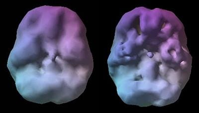
This series of articles should be particularly believable because it’s going to have some pictures of brains (but because the above assertion is accompanied by an image of a brain the citation is in the footnotes4!) 🧠
Worry Not, We’re Not Actually Going Further Than X-rays Today!
Brain Pictures5 or “neuroimaging” is of course a subset of the medical discipline of radiology, which, from it humble beginnings with x-rays has advanced through the unimaginably complex physics of magnetic resonance imaging all the way to the hybridization of computed tomography plus positron emission tomography. I’m going to guess that not everyone reading this even knows what those words mean. To be honest, I’m gonna have to look up what tomography means. Is that that T letter in PET scan? Yep:
Our first definition is from Wikipedia:
Tomography is imaging by sections or sectioning that uses any kind of penetrating wave.
So here’s how confusing this is. Even with a definition, I still don’t know what the hell it’s talking about, and given this is an article tasked with using amusing writing to convince people to read about science, I’m going to add one more definition and see if we get any closer:

Although I really thought this was going to be related to that etymology of the word tomato, it’s not. Bummer.
Let’s see if I can come up with a sentence in which this will be helpful:
Computed tomography is using x-rays and a computer to make pictures of your body, rendered in slices, section by section.
This way, were able to take what is a bunch of x-ray data and turn it into useful pictures?
I think I might’ve nailed it.
Empiricism6, demonstrated right here in the article itself.
Let’s explain a few more types of imaging just so all on the same page:
Radiographs, also known as “x-rays”
X-ray: x-rays, or “radiographs” use really high energy photons, that’s the same thing they were talking about when we’re talking about Light. Photons can be Red, photons can be Blue, photons can be really low power radio waves, and they can be really high power x-rays. When you take a really high power x-ray, which is a photon vibrating very very fast, with a higher frequency than even ultraviolet light, it can penetrate through the human body. Some of those x-rays will get absorbed on the way through, and others will not.
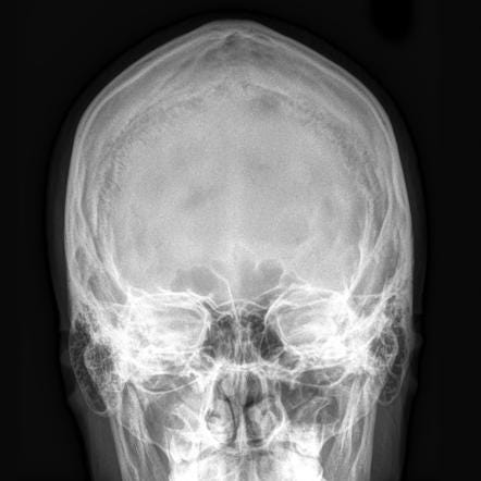
What determines whether they will go all the way through, and eventually hit a photographic plate on the other side?7 Well, if they hit something dense they’re more likely to be absorbed. If those photons hit something less dense, they cruise through. Bones are dense. Thus they are white on an x-ray because all those x-ray photons got absorbed and didn’t go through to the photographic plate on the back. When passing through the body containing muscle and blood vessels and brains and stuff, less of them get absorbed, and more of those photons hit a photographic plate. Like visual spectrum photons hitting a film negative, it gets darker. When we see x-ray radiographs, we’re looking at negatives, and unlike in photography, we don’t bother to flip the image so that white gets dark to the eye.
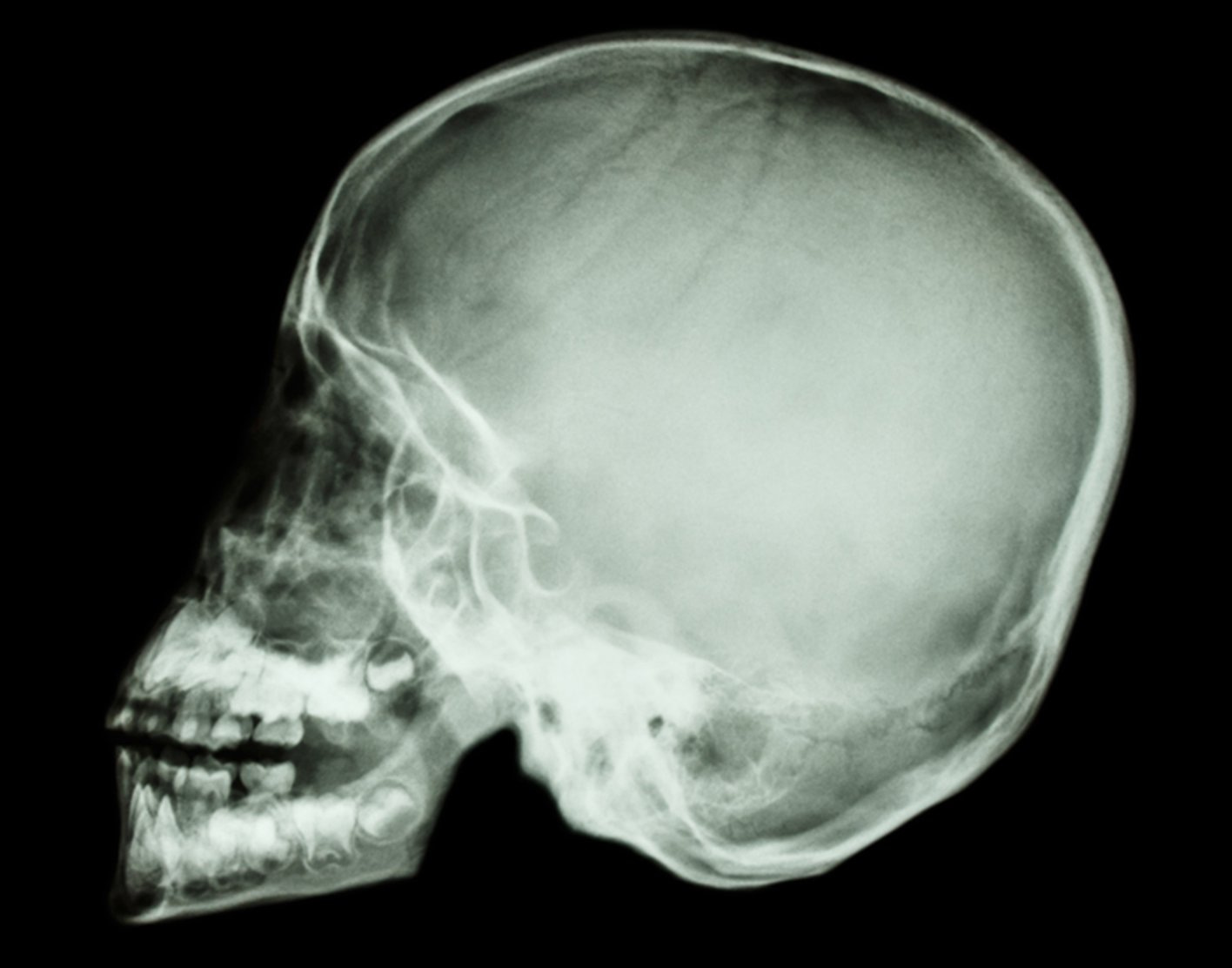
But that’s how we get an x-ray machine to make an x-ray picture. And then doctors can look at that and see what’s inside your body. We get the best resolution of bones — they’re the most dense.
Computed tomography, or CT scan
Computed tomography, as referenced above, takes the same x-rays, but uses a hell of a lot more of them. Keep in mind high frequency, high energy radiation, which is what an x-ray is, can give you problems if you get too much of it. But will get to that a little bit later.
Computed tomography takes a spinning x-ray camera, you were in the center of the tube, and you move through it, and there’s a camera spinning that you can’t see inside it shooting x-ray photons at your body and capturing them on the other side, because there’s a spinning digital film plate that you also can’t see inside the computed tomography machine!
If it made a spinning noise when you were inside, that was actually spinning you were hearing. What was spinning was the “camera” and the “photographic plate,” in X-ray digital format. There is obviously physics to how this all works, and then there’s a bunch of mathematics that goes on to take those thousands of captured little mini x-rays and turn them into slices of your body visually so doctors can casually scroll through and see everything that’s going on inside you, at least if it’s the kind of everything that can absorb an x-ray.
CT is not the most high resolution we can get, but taking a computer plus x-rays equals the ability to make slice by slice pictures of your body to see what doctors need to see, sometimes.
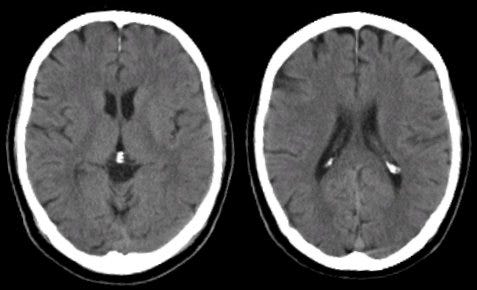
The downside is radiation. This is a risk if you get too much of it. The upside is now health professionals can see what’s going on inside you, and if it’s a big deal like bleeding, act faster. Bleeding shows up really well because it’s dense, and thus bright white on the scan. Bone fractures are also well visualized. Bone also is bright white, but not looking like a regular unbroken bone, you get the idea.
The resolution isn’t that good. It’s good for things that need to be done fast, or cheap, or especially, both. It’s almost useless for imaging the brain, unless we’re trying to see if there’s bleeding, or midline shift, or something really obvious. If there’s bleeding, it’s awesome.
If you need to look fast, because you’re worried someone had a stroke and it might be hemorrhagic, which means bleeding, a computed tomography scan will save your life. It’s good at big obvious life-threatening changes, and it’s fast, so we can do it easily in the ER.
It also has a role in acute stroke, again because of the speed of the scan, and the ability to do some fancy math when it comes to perfusion of the brain with blood, if we suspected stroke. When every minute counts, the CT scan is where it’s at. If you add contrast— more dense stuff to increase the ability to catch x-rays in soft tissue— it can do other things like pick up blood clots in the lungs, or problems in your abdomen.
Here’s a quick video that’s a nice guide to how to read a head CT scan if you ever need to understand how that’s done:
Editorial note: the following YouTube video has been pointed out to be incorrect. Thank you neuroimaging professionals!
Fluoroscopy
So one static x-ray picture is a radiograph. A movie taken with an x-ray camera is called fluoroscopy. It’s a real time visualization, usually in the process of doing a procedure, by which a doctor can watch what’s happening inside the body, particularly if they’re sticking a needle or wiggling a wire around through a blood vessel or something, and they do this by having a bunch of x-rays shooting at the person and picking those up on the other side so they get this pretty basic movie of what’s going on. The video camera of x-ray land. These are high energy photons, and there are more of them than just one picture, so this can be risky from an x-ray exposure perspective. But if you’re having a procedure, it’s helpful to have a film to guide it. That’s what fluoroscopy does.
X-ray angiogram
X-ray angiogram of the head is another x-ray, modality for head imaging, and is often useful and the diagnosis of conditions like moyamoya disease. We stick radio opaque dye into the blood, and get images like this:

You can really see the blood vessels. Clearly thanks to the dye that absorbs x-rays. These images are presented as a “positive”, so blood + dye is black, not white, as usual.
Coming up next!
In the next installment of this, we will review in common language magnetic resonance imaging. I’m not gonna drown you in math either. This will also include information about functional magnetic resonance imaging!
After that, we will get two more esoteric scans like PET, FDG-PET, and even SPECT. This is, of course, the favorite modality of Dr. Daniel, Amen, which was referenced above in my humorous parody video, and has been called out as an area of ethical concern in the field of psychiatry. As we all know now thanks to Experimental History the peer review process has failed us miserably, and that includes all the journals that publish the theoretical peer reviewed research from hucksters.
We will examine the evidence, and you can make up your own mind! I am not remotely a radiologist, so, if you are one, or a physicist, feel free to correct anything I have misunderstood!
—Owen Scott Muir, M.D.
Yes I am thinking of someone specific. No I’m not gonna make an enemy by naming them. Yes it’s you think it is.
If insecurity is not a great reason to do things I’m going have to take back everything I’ve said about Mark Cuban, and cost plus drugs, to whom I owe an apology!
My General Psychiatry residency graduating class of 2015 at The Zucker Hillside Hospital included three male residents of South Asian decent: Navin Dargani, M.D., Padam Bhatia, M.D., and Deepak Sarpal, M.D.. As a feature of the casual racism to which they were subjected, there was a certain member of the administration who could not tell them apart. And thus this human just referred to them all as Navin. As a class we started referring to them as the Navins, with their assent, in grim solidarity. They subsequently started referring to each other as Navin. As in:
Padam and Deepak at lunch:
Padam: “Hey, Navin”
Deepak: “sup”
Padam: “have you heard from Navin?”
Deepak: “yeah, I think he’s coming to class after call tomorrow anyway”
Padam: “dedication man. Hey, Navin?”
Deepak: “yeah, Navin?”
Padam: “can you grab me a coffee before class, need to go answer this page”
Deepak: “Navin, anything for you”
Padam, on phone: “this is Dr. Navin, how can I help you?”
Etc.
And it basically continues to this day. As a public service announcement I will remind readers: “I can’t tell you brown people apart” is neither funny, cute, nor professional. Learn the names of people even and especially if they are hard to say or spell and/or not white or male.
So many papers on this topic, here are a few winners:
https://pubmed.ncbi.nlm.nih.gov/17803985/
Gruber, D., & Dickerson, J. A. (2012). Persuasive images in popular science: Testing judgments of scientific reasoning and credibility. Public Understanding of Science, 21(8), 938–948. https://doi.org/10.1177/0963662512454072
Wouldn’t it be funny if we called then brain pictures? it’s the kind of thing doctors would never do because it doesn’t line up with our whole ego thing about speaking in entirely different language based on Latin when we’re talking about medical stuff so that people think we’re smart. The ego!
Empiricism is the approach to life in which questions that could be questions are actually answered. The dictionary definition is as follows:
em·pir·i·cism
noun PHILOSOPHY
the theory that all knowledge is derived from sense-experience. Stimulated by the rise of experimental science, it developed in the 17th and 18th centuries, expounded in particular by John Locke, George Berkeley, and David Hume.
I did pretty good with that. I didn’t reference David Hume, so not quite as well as I could’ve done, but once were talking about John Locke and David Hume, we’re pretty sure that this is some high-end shit. I mean, I clearly went to college. And now you feel like you’re able to impress your friends with philosophical chit chat too. Your next companion at dinner has my sincerest apologies, because you’re going to sound pretty smug.
The is the x-ray film that we later develop and view back in the day, and these days it’s done digitally.
In case people didn’t catch it, that video is a parody. It was done for an event where people had to guess whether the information was real or fake, and I am not a consultant to the amen clinics. That was for the purpose of fakeness, and people had to guess if it was real.